 Pre-commercial · patents pending
Equine Mosaicplasty — Osteochondral Grafting System for Subchondral Bone Cysts
- 31 cases (1998–2023)
- 90% lameness improvement
- 68% returned to full athletic performance
Overview
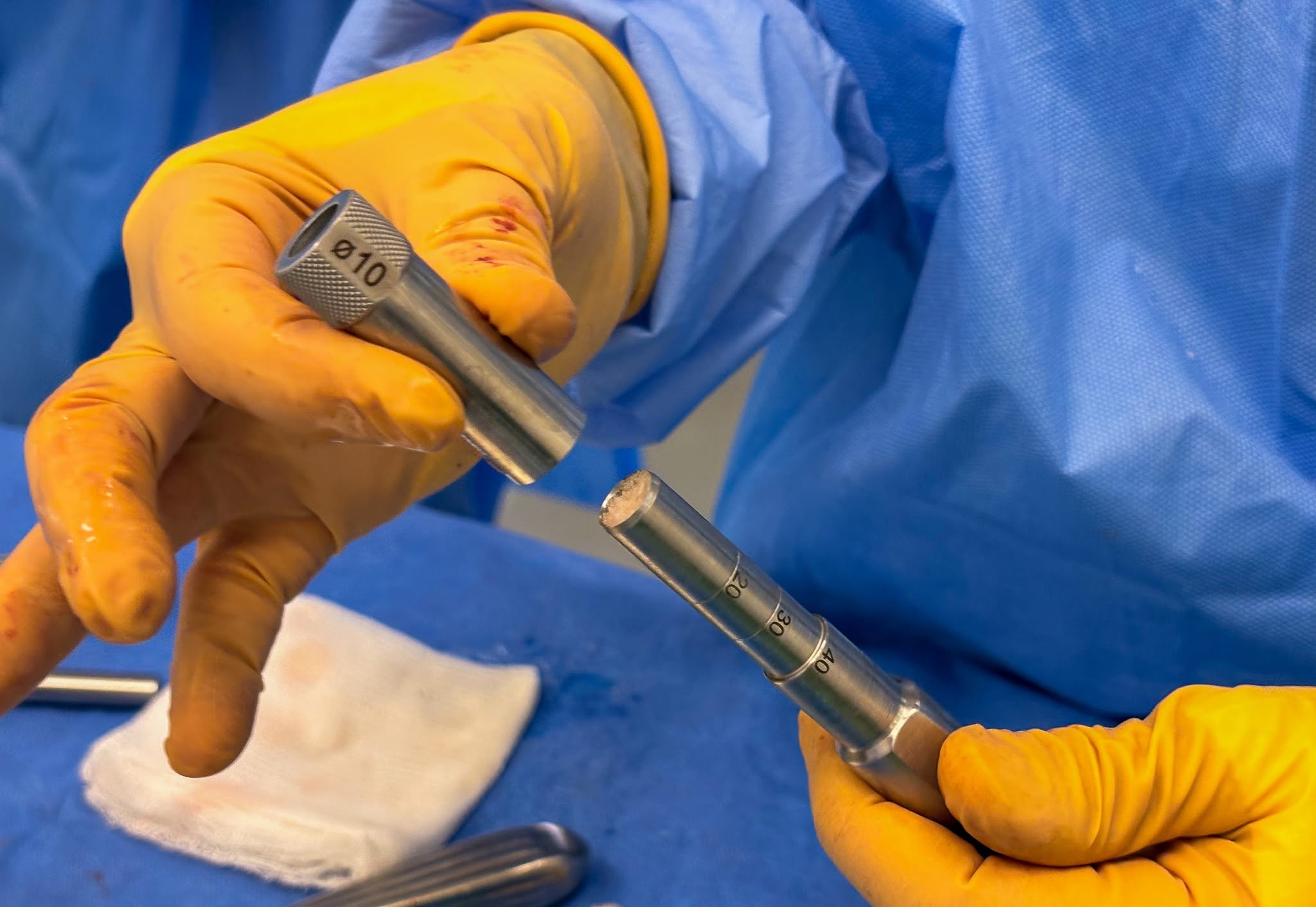
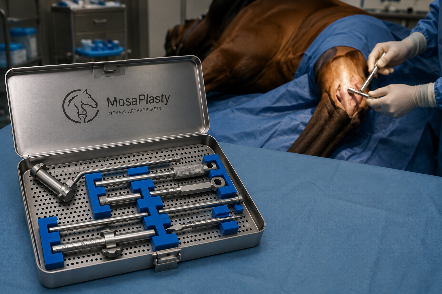
Subchondral bone cysts are a common cause of lameness in performance horses, and existing treatments show inconsistent long-term results. Vet-Inno TTC’s mosaicplasty technique offers a single-step solution: cylindrical bone grafts are harvested from a low-weight-bearing joint surface and press-fit into the cyst using a custom-designed instrument set built specifically for equine anatomy — unlike adapted human orthopedic tools, which lack the necessary length and durability for horses.
The technique’s long-term effectiveness is backed by 25 years of clinical use and a peer-reviewed retrospective study of 31 cases treated between 1998 and 2023: most horses regained soundness and returned to full athletic performance, with favorable outcomes across a wide age range — including mature horses, where other techniques typically perform less well.
Evidence & results
Source: Pál Z, Tuska P, Vásárhelyi G, Hangody L, Hurtig M, Kaposi AD, Bodó G. “Mosaic arthroplasty in equine stifle and fetlock joints: A retrospective study of 31 cases between 1998 and 2023.” Veterinary Surgery, 2025. DOI: 10.1111/vsu.14296 (open access).
- Multi-institutional retrospective study, 31 horses treated across four institutions between 1998 and 2023, minimum 12-month follow-up (median 24 months, up to 10 years)
- 90% of horses showed improved lameness after surgery; 68% returned to the same or higher level of athletic performance
- Outcomes held up in mature horses, not just young ones — a point of difference from other published techniques, which tend to report success mainly in 2- and 3-year-olds
- Horses receiving fewer grafts (≤2) trended toward better outcomes than those needing 3 or more
- Complication rate was low (16%, 5/31), and most complications were minor and resolved with routine treatment
- The study also documents the 25-year evolution of the custom instrument set and surgical technique, including refinements that reduced graft-insertion errors
What we’re looking for
We’re looking for two kinds of partners: equine clinics interested in offering this technique, and a reseller/distributor for the instrument set.
Request more informationSuccess story
Lenny: a demonstration surgery at Colorado State University
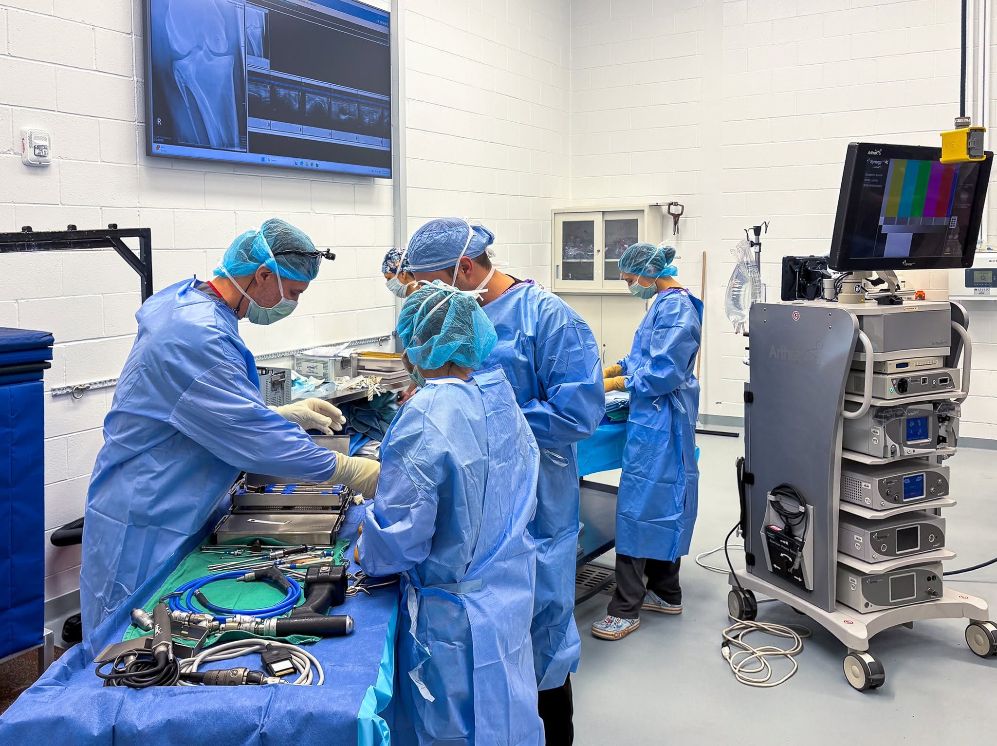
In spring 2026, Prof. Gábor Bodó — head of the Department and Clinic of Equine Medicine, and the person who adapted this technique for equine use (the original human-orthopedic mosaicplasty method was developed by Prof. László Hangody) — was invited to Colorado State University to perform a demonstration surgery. The invitation came from Prof. Laura Goodrich, director of CSU’s Research Center and a former president of the American College of Veterinary Surgeons (ACVS), recognized as one of the leading US experts in equine joint-surface reconstruction.
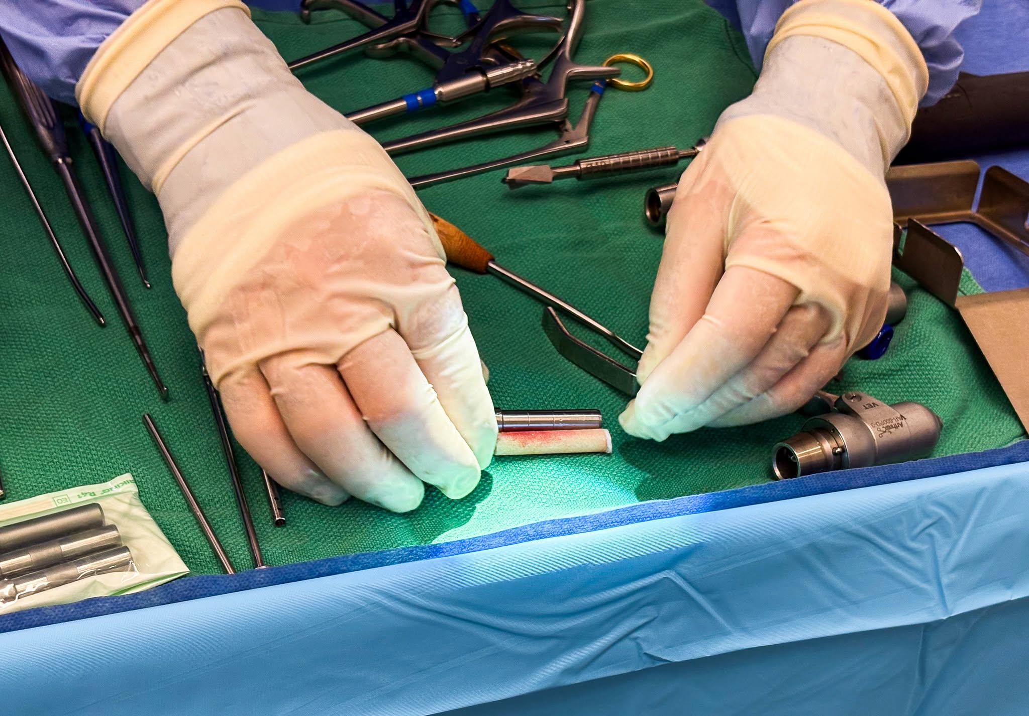
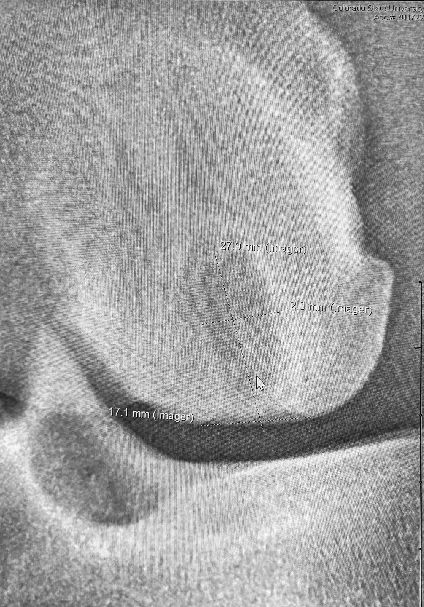
The patient was Lenny, a 4-year-old Quarter Horse with knee pain whose owner hoped for a future barrel-racing career. Imaging revealed a large subchondral bone cyst in the medial femoral condyle — a case the Colorado team judged best suited to the Hungarian technique. Prof. Bodó implanted three osteochondral grafts (10 mm, 8.5 mm, and 6.5 mm in diameter, 30 mm long) to fill the defect with the horse’s own healthy, cartilage-covered bone.
Recovery has gone well: Lenny is sound, bearing weight normally, and progressing through a structured rehabilitation program (swimming, massage, and a gradually increasing exercise load) aimed at a return to competitive form. His owner has reported being very satisfied with the results so far. As Prof. Bodó put it, Lenny’s recovery could not have gone any better — and this same sling-assisted recovery technique is also available at the university’s own Üllő clinic.
Source: Lóklinika (Equine Clinic, University of Veterinary Medicine Budapest) Facebook page